Injury Prevention

Frozen Shoulder: Unravelling the Complexities and Providing Clarity

Written by Michael Crawley, BSc, BPT, CSCS

Nearly 100 years ago, Earnest Codman coined the term “frozen shoulder” and highlighted three clinical issues (Salamh et al. 2025):

  • Difficult to define

  • Difficult to treat

  • Difficult pathology to explain to patients

Those three points still hold true today.

Multiple structures and pathological findings have been implicated in the development of frozen shoulder. This includes the accumulation of immune system mediators, thickening of ligaments, and altered collagen translation (Pandey and Madi 2021). Clinically, this presents as a shoulder with reduced range of motion in both active and passive flexion, abduction, and external rotation (as seen in the image below).

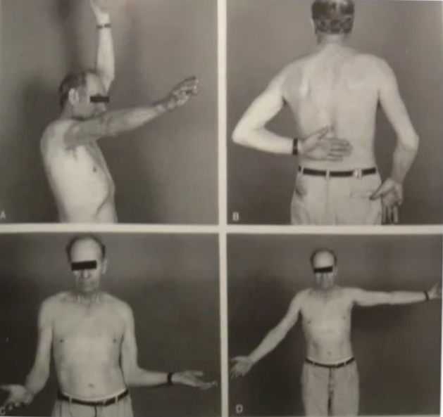
Figure 1: Reduced Shoulder range of motion (ROM) with frozen shoulder

The Real Impact of Frozen Shoulder

A scoping review examining how people experience and live with frozen shoulder demonstrates how debilitating and impactful the condition can be. King and Hebron (2023) identified five major themes:

  1. “Dropping me to my knees, due to the pain”

  2. Struggle for normality

  3. Emotional change for self

  4. Challenges through the healthcare journey

  5. Coping & adapting

This highlights that frozen shoulder is not just a physical limitation. It can significantly alter how someone functions and experiences their daily life.

Unfortunately, frozen shoulder demonstrates a bias towards a particular demographic. Females in the 40–60 age category take the brunt of diagnoses. To rub salt in the wounds, females are more likely to experience a more prolonged and symptomatic course compared to male counterparts.

Types of Frozen Shoulder

Frozen shoulder can be broadly classified into two categories (Pandey and Madi 2021):

Primary:
A stiff shoulder developing with no known cause. However, there are commonly linked conditions, most notably diabetes mellitus and thyroid dysfunction. The incidence of frozen shoulder can reach as high as 30% in individuals with diabetes.

Secondary:
A stiff shoulder with an underlying cause such as direct trauma (e.g. a fall), infection, or inflammatory conditions.

The Three Stages of Frozen Shoulder

Frozen shoulder follows a series of stages, delineated by changing symptoms (Date and Rahman 2020). While approximate timelines are often attached, there is significant variability, and for some individuals, full resolution may not occur within 3–5 years.

Freezing Stage (Stage 1: 2–6 months)

  • Predominantly characterised by moderate to severe pain and partial restriction of ROM

  • Early stages may present with pain and only terminal loss of ROM

This stage can be confused with rotator cuff tendinopathy. However, ROM does not progressively worsen in tendinopathy, whereas it continues to worsen with each follow-up in frozen shoulder.

Frozen Stage (Stage 2: 4–12 months)

  • Characterised by both pain and stiffness in varying proportions

  • Early phase tends to be more pain-dominant

  • Later phase becomes more stiffness-dominant

Thawing Stage (Stage 3: 6–26 months)

  • Characterised by minimal pain

  • Gradual resolution of stiffness

  • Progressive return of movement

Pathologically, this reflects a gradual reduction in inflammation and restoration of movement.

Treatment and Management Across the Stages

What actually works, and when it matters

The research on the effectiveness of treatments for frozen shoulder remains conflicting. However, a conservative approach is typically recommended as the starting point (Date and Rahman 2020).

Common interventions include:

  • Analgesics

  • Physiotherapy

  • Intra-articular injections

  • Suprascapular nerve block

Early Stage: Movement Within Tolerance

In the early stage of frozen shoulder, gentle stretching and mobility exercises within a pain-free range are advised (Date and Rahman 2020).

Creativity can play a key role here, as Louis Gifford, the brilliant pain specialist, stresses. In his book Aches and Pains, he explains how adjusting body position can influence the amount of pain-free range available to a limb.

The videos below demonstrates this concept. The key idea is simple:

  • The arm can move relative to the body

  • Or the arm can stay fixed while the body moves around it

Shoulder Range of Motion Wall Drills:
https://youtu.be/9_GwO7r24hM

Passive and active-assisted exercises can also be incorporated. These reduce the working stress on affected structures, allowing the humerus to move through range without generating or exacerbating pain.

Active Assisted and Passive Shoulder:
https://youtu.be/072jZDVW-ac

As Pain Settles: Introducing Strength

As pain begins to reduce and become more manageable, strengthening exercises can be introduced.

Here, the principle that “the dose and position make the poison” becomes particularly relevant.

Using isometrics in varying positions and directions allows for global loading through the shoulder while staying within tolerable limits.

Entry Level Isometric:
https://youtu.be/mDzgyyKlzZo

Later Stages: What Are Mobilisations Actually Doing?

Mobilisations performed by a physiotherapist in the later stages have shown some utility. However, the mechanism behind their effectiveness is contested.

For many years, the prevailing thought was that inferior mobilisation directly impacted the shoulder joint capsule. However, Jeremy Lewis, a well-known Australian shoulder specialist, has pointed out that a physiotherapist would need to generate approximately 600kg of force to meaningfully affect the capsule.

I am not aware of many Canadians with a 600kg deadlift.

The best approach at this stage would be to continue to progress strength training through pain free range.

Injections and Medical Management: Timing Is Key

Outside of physiotherapy, injections and pharmacological treatments are often used.

Nonsteroidal anti-inflammatories have shown little impact in the case of frozen shoulder. Intra-articular steroid injections, however, have demonstrated positive effects, particularly when used at the right time.

Again, Jeremy Lewis stresses that these injections must be used in the early stages, when pain is highest. This reinforces the importance of early and accurate diagnosis.

A similar pattern is seen with suprascapular nerve blocks, which can also have a positive effect on pain relief when applied early (Date and Rahman 2020).

Surgical Options: Often Less Helpful Than Expected

Surgical options are available, but often yield little additional benefit.

Beard et al. (2018) found no clinically significant benefit of shoulder arthroscopy compared to sham surgery. This was further supported by the large UK FROST trial (Corbacho et al. 2021), which reported that early physiotherapy was more cost-effective and accessible compared to invasive and costly surgical approaches.

Interestingly, manipulation under anaesthetic, which previously had negative connotations, has shown some efficacy. This likely relates to the reduction of muscle guarding and tension that can develop with frozen shoulder. When under anaesthetic, this guarding effect is temporarily removed.

Looking Beyond the Shoulder

An important point that is often not expressed or evaluated in the research is that frozen shoulder may be a sign of broader health issues, stemming from multiple systems in the body.

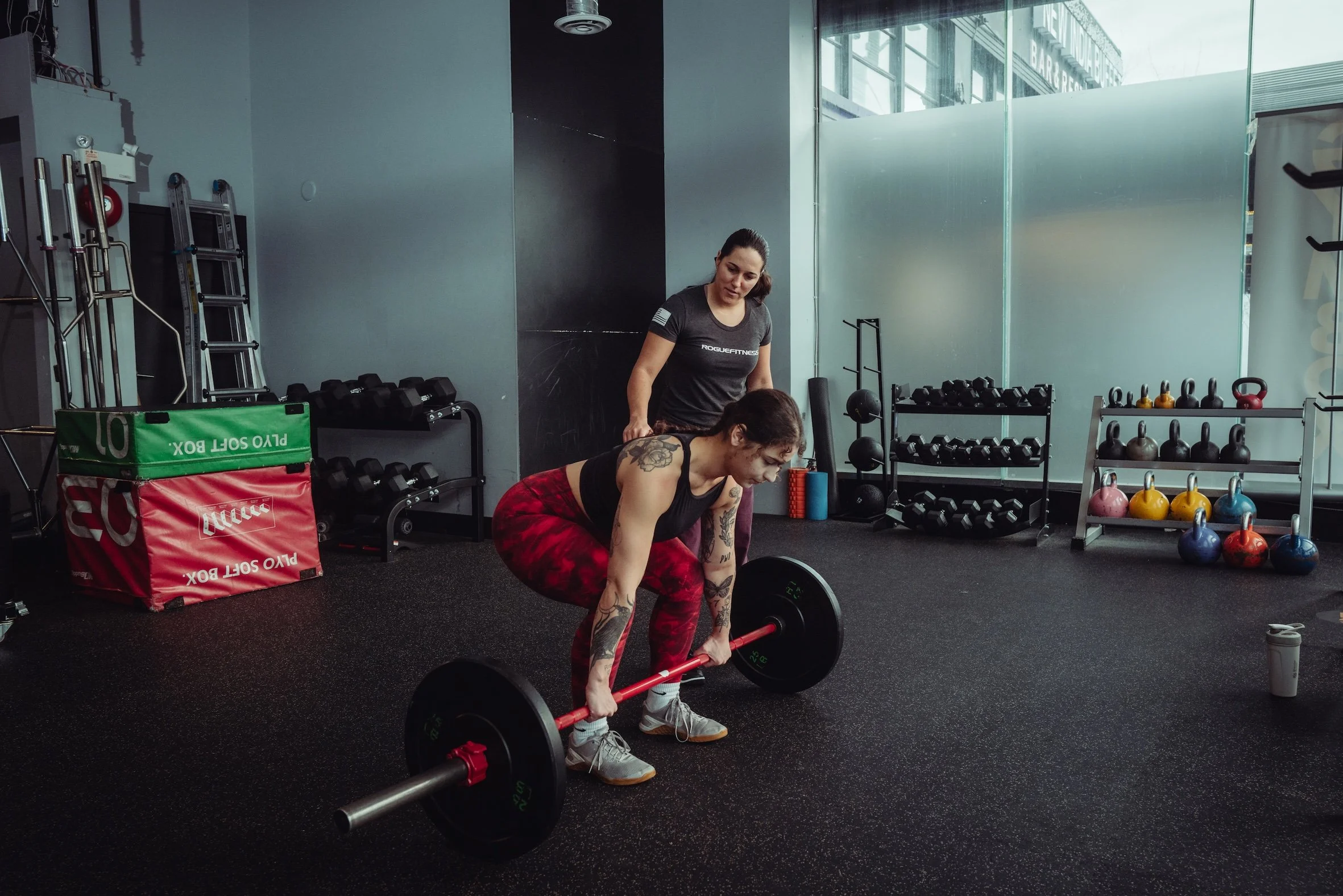
In many cases, it can act as a wake-up call to incorporate strength and conditioning into your lifestyle and address other health metrics.

You may not be able to train the affected side in the same way, but there are still many full-body exercises that can be performed without exacerbating the shoulder:

  • Towing a sled

  • Belt squat

  • Walking lunges

  • Step-ups

Why This Matters

There are three key reasons why this approach is important:

  1. Approximately 1 in 5 people go on to develop similar symptoms in the opposite shoulder (Pandey and Madi 2021)

  2. Sedentary individuals are more likely to receive a frozen shoulder diagnosis

  3. Well-designed strength and conditioning programs can positively influence the systems linked to frozen shoulder development, including endocrine, immune, and cardiovascular systems

Deeper Dive into Causation and Management

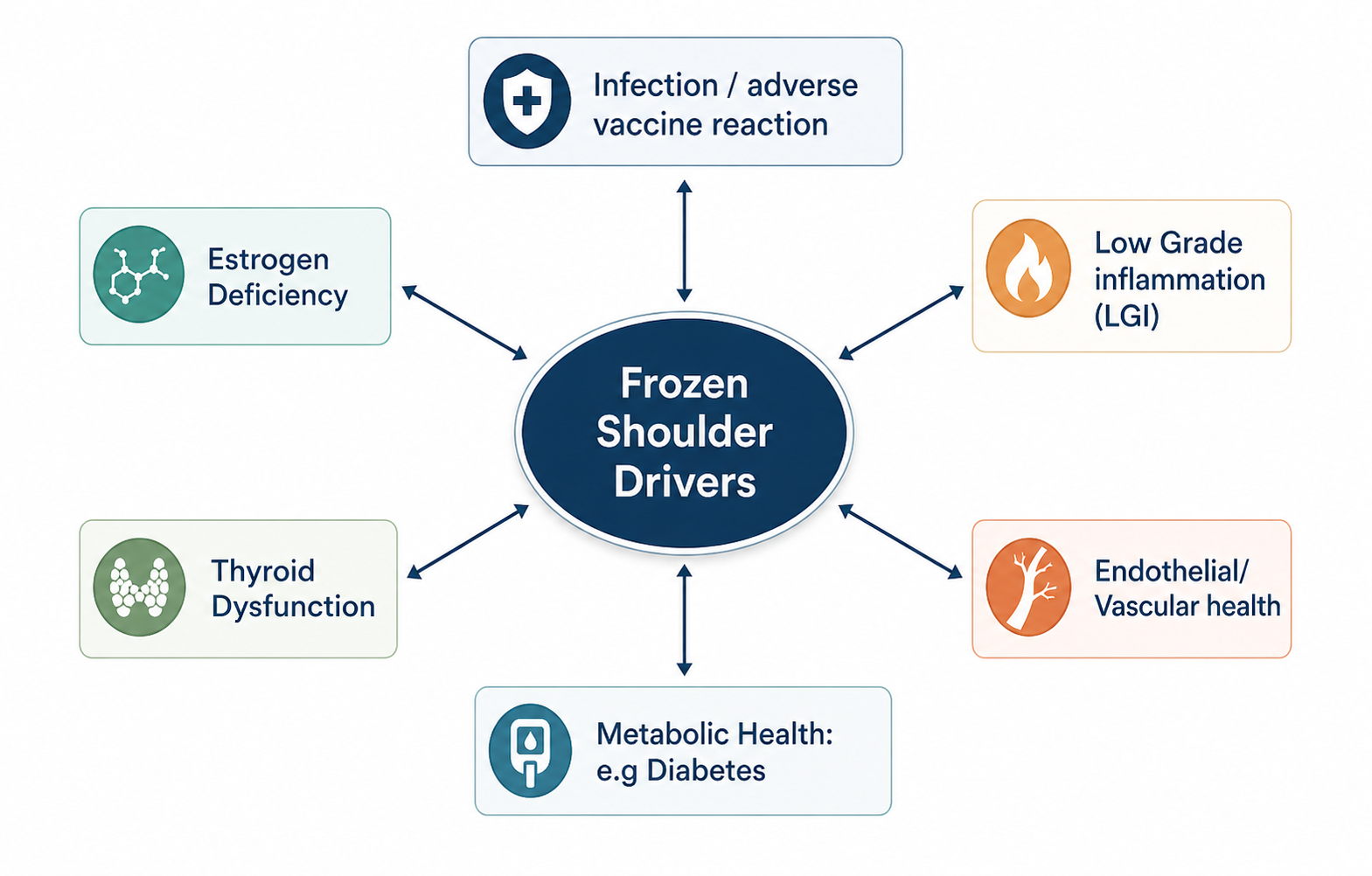
Recent research has continued to highlight the multi-faceted nature of frozen shoulder and the challenges associated with its management (Navarro-Ledesma 2025a).

This is not a condition driven by a single structure or isolated tissue. Instead, it reflects the interaction of multiple systems within the body.

The diagram below highlights this well. Rather than being caused by one specific issue, frozen shoulder appears to sit at the intersection of several physiological systems, all of which can influence one another.

Estrogen and Menopause

One of the more consistent patterns seen in the research is the increased prevalence of frozen shoulder in peri-menopausal women. This has led to estrogen being identified as a key player in its development (Wend et al. 2012).

As shown in figure below, estrogen has effects that extend well beyond the reproductive system. Its influence spans multiple systems that are directly relevant to frozen shoulder.

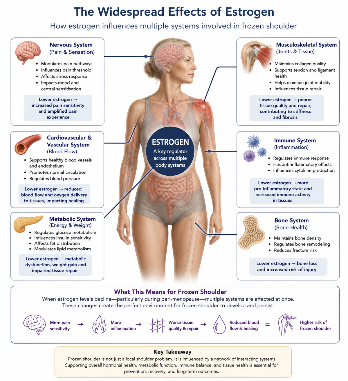
Neuroendocrine System

Declining estrogen levels can influence the nervous system through several mechanisms, impacting pain thresholds, resilience to stress, and central sensitisation.

A useful way to think about this is the “fire alarm” analogy.

You leave the bacon on the grill too long and the fire alarm goes off because of the smoke. There is no fire, but the system reacts as if there is.

With reduced estrogen levels, the threshold for triggering that “alarm” can become lower. The result is an amplified pain experience, even when the underlying tissue irritation may not fully justify it.

Metabolic System

Estrogen also plays a key role in fat metabolism, glucose regulation, and resistance to oxidative stress.

When these systems are disrupted, it can create an internal environment where tissue repair is compromised. This contributes to fibrosis, which is a hallmark of frozen shoulder.

Immune System

The same pattern continues within the immune system.

Declining estrogen levels tend to promote a more pro-inflammatory state. Immune system mediators accumulate within the tissues involved in frozen shoulder, and when combined with metabolic dysfunction, this can further drive the condition.

Targeting the System, Not Just the Shoulder

The research highlights how frozen shoulder is influenced by multiple systems, not just the shoulder itself. As a result, management is not limited to physiotherapy or surgical intervention alone.

There are a number of factors that could be explored here, but for the purpose of this piece, three of the more relevant and actionable areas will be discussed below.

Strength and Conditioning

Well-designed and properly implemented strength and conditioning programs have demonstrated positive impacts on estrogen levels, muscle mass, and fat mass in menopausal women (Razzak et al. 2019).

As mentioned previously, even with an impacted and painful shoulder, this does not mean avoiding training altogether or waiting for full resolution before doing anything.

The whole-body and multi-system benefits of strength training can influence long-term outcomes indirectly. While the shoulder itself may be limited, the broader physiological adaptations still matter.

Nutrition

Diet quality also plays a meaningful role.

A nutritional approach centred around higher-quality, minimally processed foods has been shown to impact symptom severity in individuals with frozen shoulder (Hamed-Hamed et al. 2026).

In practice, the decision to implement a structured strength training program often leads to improvements in other lifestyle behaviours, including dietary choices.

In the same way that hormonal, metabolic, and immune factors can drive the development of frozen shoulder, lifestyle decisions can push back against these drivers. This not only has the potential to improve current symptoms, but also to reduce the likelihood of future development.

Sleep and Circadian Rhythm

Circadian rhythm and sleep regulate inflammatory processes, hormonal release, and tissue repair (Navarro-Ledesma 2025a).

These are central to both general health and the development and recovery of frozen shoulder, as well as adaptation to strength training and exercise.

This is where the entanglement of systems becomes more apparent.

Bringing It Together

Sleep, exercise, and nutrition can be thought of as a three-legged stool. Each supports the others, and removing one weakens the entire system.

Addressing these factors will not provide an immediate solution to frozen shoulder. However, they can set the conditions for recovery and reduce the likelihood of recurrence, particularly when considering that approximately 20% of individuals will experience similar symptoms in the opposite shoulder.

Summary and Takeaways

Frozen shoulder is a systems issue, not just a joint problem

Frozen shoulder is not a local condition. It can have significant and long-term effects on both physical and psychological well-being.

In some cases, it can be so debilitating that it alters how an individual functions day to day. That may sound hyperbolic, but when revisiting the five themes outlined earlier, alongside the number of systems involved, it becomes more understandable.

Approaching treatment with a reductionist lens, relying solely on an injection or a home exercise program, is akin to using a hammer where a scalpel is required. This sentiment is supported in a recent review by Brindisino et al. (2026).

Effective management requires a more nuanced and personalised approach that considers the multiple drivers involved:

  • Hormonal (endocrine)

  • Immune system (autoimmune / inflammatory)

  • Strength, mobility, and capacity

  • Cardiovascular health

  • Pain psychology (sensitisation and emotional drivers)

  • Structural factors

  • Circadian rhythm and sleep

Key Takeaways

  • General strength training can still be completed and is beneficial with a frozen shoulder diagnosis

  • Surgical interventions are often unwarranted and do not demonstrate superior outcomes

  • Frozen shoulder is multi-factorial, and lifestyle factors such as exercise, nutrition, and sleep play a critical role in both management and risk reduction

References

Beard, D. J. et al. 2018. Arthroscopic subacromial decompression for subacromial shoulder pain (CSAW): a multicentre, pragmatic, parallel group, placebo-controlled, three-group, randomised surgical trial. The Lancet 391(10118), pp. 329-338. doi: 10.1016/S0140-6736(17)32457-1

Brindisino, F. et al. 2026. Beyond the capsule: an integrated perspective on the wide world of frozen shoulder. A collaborative viewpoint. Pain Management, pp. 1-20. doi: 10.1080/17581869.2026.2636725

Corbacho, B. et al. 2021. Cost-effectiveness of surgical treatments compared with early structured physiotherapy in secondary care for adults with primary frozen shoulder : an economic evaluation of the UK FROST trial. Bone Jt Open 2(8), pp. 685-695. doi: 10.1302/2633-1462.28.Bjo-2021-0075.R1

Date, A. and Rahman, L. 2020. Frozen shoulder: overview of clinical presentation and review of the current evidence base for management strategies. Future Sci OA 6(10), p. Fso647. doi: 10.2144/fsoa-2020-0145

Hamed-Hamed, D. et al. 2026. Impact of nutritional profile on pain and functionality in patients with frozen shoulder: a cross-sectional observational study. Frontiers in Medicine Volume 13 - 2026,  doi: 10.3389/fmed.2026.1785577

King, W. V. and Hebron, C. 2023. Frozen shoulder: living with uncertainty and being in “no-man’s land”. Physiotherapy Theory and Practice 39(5), pp. 979-993. doi: 10.1080/09593985.2022.2032512

Navarro-Ledesma, S. 2025a. Frozen Shoulder as a Systemic Immunometabolic Disorder: The Roles of Estrogen, Thyroid Dysfunction, Endothelial Health, Lifestyle, and Clinical Implications. J Clin Med 14(20),  doi: 10.3390/jcm14207315

Navarro-Ledesma, S. 2025b. Frozen Shoulder as a Systemic Immunometabolic Disorder: The Roles of Estrogen, Thyroid Dysfunction, Endothelial Health, Lifestyle, and Clinical Implications. Journal of Clinical Medicine 14(20), p. 7315. 

Pandey, V. and Madi, S. 2021. Clinical Guidelines in the Management of Frozen Shoulder: An Update! Indian J Orthop 55(2), pp. 299-309. doi: 10.1007/s43465-021-00351-3

Razzak, Z. A. et al. 2019. Effect of aerobic and anaerobic exercise on estrogen level, fat mass, and muscle mass among postmenopausal osteoporotic females. Int J Health Sci (Qassim) 13(4), pp. 10-16. 

Salamh, P. et al. 2025. An international consensus on the etiology, risk factors, diagnosis and Management for individuals with Frozen Shoulder: a Delphi study. J Man Manip Ther 33(4), pp. 309-320. doi: 10.1080/10669817.2025.2470461


Wend, K. et al. 2012. Tissue-Specific Effects of Loss of Estrogen during Menopause and Aging. Frontiers in Endocrinology Volume 3 - 2012,  doi: 10.3389/fendo.2012.00019

You Should Not Get Injured During a Training Session

Written by Evelyn Calado, MKin, CSCS, RKin

If you leave a training session injured, something has gone wrong.

That is not normal. It should not be expected. And it is not part of “training hard.”

I have been in this industry for over a decade, and I have seen far too many situations where clients get hurt in the weight room. Not because of bad luck, but because of poor decisions.

The Gym Should Be One of the Safest Places You Train

Think about it.

The weight room is a controlled environment.

  • You control the load

  • You control the movement

  • You control the pace

  • You control the rest

Compare that to sport, where there are opponents, unpredictable movements, and variables you cannot control.

In theory, your injury risk in the gym should be extremely low.

That does not mean training is easy. You should still be challenged. You should still push your limits.

But it should be done in a controlled and intentional way.

Where Things Go Wrong

Most training-related injuries are not random. They come from avoidable mistakes.

1. Ego-Based Training

This is one of the biggest issues.

A client walks in, maybe they look strong or athletic, and the session becomes about proving something. The load goes up too quickly, technique breaks down, and fatigue is ignored.

That is how people get hurt.

2. No Plan

This is more common than people think.

Clients come in and have no idea what they are doing that day. The trainer is choosing exercises on the spot with no structure, no progression, and no record of previous sessions.

Without a plan, there is no progression. Without progression, there is no direction. And without direction, you are just accumulating risk.

3. Poor Exercise Sequencing

Fatigue matters.

If someone is pushed to the point of exhaustion through their legs and core, and then asked to perform a heavy compound lift, that is a problem.

That is not “hard training.” That is poor decision-making.

4. Ignoring the Individual

Not every client should move the same way.

Mobility, injury history, movement patterns, and training experience all matter. If those are ignored, you are forcing someone into positions they are not prepared for.

That is where breakdown happens.

Soreness Is Not the Goal

Another misconception is that a good session should leave you unable to move.

If your trainer’s goal is to completely destroy you, that is a red flag.

  • You should not struggle to sit at your desk

  • You should not be unable to walk for days

  • You should not feel worse instead of better

Anyone can make you tired.

It takes skill to build a program that challenges you, progresses you, and still allows you to function.

What Good Training Actually Looks Like

Good training is not random. It is structured.

  • There is a clear plan

  • There is progression over time

  • Your loads and performance are tracked

  • Exercises are selected for a reason

  • Intensity is managed, not guessed

You are pushed, but within your capacity.

You improve, without being completely broken down in the process.

Playing the Long Game

At Avos Strength, the focus is simple:

Train. Play. Repeat.

The goal is not to win a single session. The goal is to keep you training consistently, improving over time, and continuing to do the things you enjoy.

That means:

  • Checking your ego at the door

  • Building progressively

  • Respecting your current capacity

  • Training with intention

The Bottom Line

The gym should not be where you get injured.

Can things happen occasionally? Yes. But injuries should be rare, not expected.

If you have worked with a trainer and felt unsafe, unsupported, or left sessions worse than when you walked in, that is not something you should accept.

You deserve better coaching than that.

The Limb Arc Model: Why You Should Train the Range of Motion You Actually Own

Written by Evelyn Calado, MKin, CSCS, RKin

If you’ve ever wondered why:

  • Your knees cave in at the bottom of a squat

  • Your low back extends when the weight gets heavy

  • One hip always feels “stuck” at 90°

  • Or mobility drills don’t seem to transfer to strength

…you’re probably running into a concept explained by the Limb Arc Model.

This model, commonly attributed to Bill Hartman, describes how rotational bias changes across ranges of joint flexion — particularly at the hip. And once you understand it, exercise selection becomes dramatically more logical.

Let’s break it down.


What Is the Limb Arc Model?

The Limb Arc Model proposes that rotational leverage changes as a joint moves through flexion.

At the hip specifically:

  • Early flexion favors external rotation (ER)

  • Mid-range flexion favors internal rotation (IR)

  • Deep flexion returns to an external rotation bias

This is not arbitrary. It reflects changes in joint geometry, length tension relationships, and moment arms.

Most people train hip flexion as if it is one continuous quality. It is not. It is three mechanically distinct regions.

That shift matters for:

  • Squats

  • Deadlifts

  • Split squats

  • Gait mechanics

  • Sport performance

  • Injury risk

The Hip Flexion Arc Explained

Here’s the simplified breakdown:

0–60° Hip Flexion → External Rotation Bias

In early hip flexion, the joint favors:

  • External rotation

  • Abduction

  • Supination at the foot

  • Sacral counternutation

In gait, this corresponds most closely with early stance, when the heel has contacted the ground and the pelvis is relatively externally rotating as load is being accepted.

In the gym, this is the top portion of a squat or the early phase of a hinge.

External rotators and abductors have favorable leverage here.

60–100° Hip Flexion → Internal Rotation Bias

Around 90° hip flexion:

  • Internal rotators and adductors have improved leverage

  • Length–tension relationships favor IR

  • The piriformis shifts moment arm toward IR

  • The sacrum moves toward nutation

  • The foot transitions toward pronation

In gait, this corresponds most closely with mid stance, when the pelvis is internally rotating on the femur and vertical ground reaction forces are highest.

In a squat, this is typically around parallel.

100°+ Hip Flexion → Returns to External Rotation Bias

As you approach deep hip flexion:

  • The system transitions back toward ER

  • Supination strategies often reappear

  • External rotators regain leverage

This helps explain why some people feel “better” deep in a squat even if they struggle at parallel. They are returning to a range where external rotation leverage increases again.


Why Internal Rotation at 90° Matters

Most loaded bilateral lower-body exercises demand control around 60–100° hip flexion.

If internal rotation is limited in that range, common compensations show up:

  • Knee valgus

  • Lumbar extension

  • Butt wink

  • Hip shifting

  • Over-pronation

  • Gripping with toes

This is not always a strength problem.

It’s often a relative motion problem.

The joint is being asked to produce force in a range it does not control. When the femur is not internally rotating relative to the pelvis, the pelvis, spine, or foot moves instead.


“Train within the Range You Own”

Here’s where this becomes practical.

Owning a range means:

  • You can access it

  • You can control it

  • You can breathe in it

  • You can maintain joint relationships without compensating

If you lack IR at 90°, loading it heavily won’t fix it.

It may:

  • Reinforce compensations

  • Drive orientation strategies (like anterior pelvic tilt)

  • Increase compressive strategies instead of restoring motion

Instead, you might need:

  • Split squats that bias mid-stance

  • Exercises emphasizing medial arch contact

  • Internal rotation control drills

  • Breathing-based repositioning work

  • Heel references to restore early stance mechanics

Force production should follow motion restoration — not precede it. Ie; Restore control first. Then add load.


How This Applies to Programming

The Limb Arc Model gives you a filter for exercise selection.

The question is not whether someone “has internal rotation.”

The question is where in the arc they lose control.

If Control Breaks Down Between 0 and 60 Degrees

You will see:

  • Difficulty accepting load at the top of the squat

  • Poor heel contact

  • Immediate external rotation gripping

  • Early lumbar extension

In this case, reinforce early stance mechanics.

Use closed chain drills that emphasize heel reference and controlled external rotation.
Keep the hip in the zero to sixty degree range and teach load acceptance without extension strategies.

The goal is stable external rotation control in early hip flexion.

If Control Breaks Down Between 60 and 100 Degrees

You will see:

  • Knee valgus at parallel

  • Hip shift at ninety degrees

  • Lumbar extension at the sticking point

  • Loss of medial arch control

This is the most common presentation.

Here, you bias time spent in sixty to one hundred degrees of hip flexion in closed chain.


Split squat variations are useful when organized correctly because they allow:

  • Pelvis on femur relative motion

  • Clear stance leg reference

  • Control of hip flexion angle

  • Moderate load that does not overwhelm internal rotation capacity

The key is managing support and load so that the pelvis can internally rotate on the femur without defaulting into orientation strategies such as anterior pelvic tilt or lateral shift.

This is not about making someone balance harder.

It is about placing them in the internal rotation biased window and allowing them to control it.

If Control Breaks Down Beyond 100 Degrees

You will see:

  • Instability or collapse in deep squat

  • Over reliance on passive structures

  • Loss of tension in the bottom

In this case, gradually expose the athlete to deeper flexion under controlled conditions, restoring external rotation leverage without compensatory lumbar flexion.


Why This Model Is Powerful

The Limb Arc Model connects:

  • Gait

  • Breathing mechanics

  • Pelvic motion

  • Squat depth

  • Performance

  • Compensation patterns

It explains why:

  • One depth feels strong and another feels unstable

  • Deep squats don’t fix mid-range weakness

  • “Mobility” doesn’t always transfer to strength

Because leverage changes as joint angles change.

And if you don’t own the transition between those zones, the body will compensate.


Final Takeaway

The Limb Arc Model isn’t about stretching more.

It’s about understanding that:

Rotational demands shift as joints move through flexion.

And if you load a range you don’t own, your body will borrow motion from somewhere else.

Train the range you control.

Then expand it.

That’s how you build durable strength.

Learn more about how we assess movement and build individualized programs at Avos Strength.


ACL Injuries: How They Occur, Who Is at Risk, and Why Training Quality Matters (Part 1)

Written by Michael Crawley, BSc, BPT, CSCS


BACKGROUND

Anterior cruciate ligament injuries (ACLI) are often viewed as sudden, unavoidable events that are “fixed” through surgery. In reality, both injury risk and long-term outcomes are strongly influenced by training quality, rehabilitation approach, and the decisions made before and after injury.

This article highlights the complexity of ACL injuries, explains how and why they occur, and outlines key training and rehabilitation considerations that influence risk and return to sport outcomes. While ACL injuries are often discussed in isolation, they are rarely simple knee injuries, and successful outcomes require a broader, long-term view.

The information presented is intended to provide practical, actionable insight for a range of athletes and stakeholders, including:

  • Youth multi-sport athletes and their parents

  • High-level collegiate and professional athletes

  • Competitive recreational athletes of all ages

ACLI have increasingly been described as an epidemic across both amateur and professional sport. Several studies report that ACL injuries account for approximately 50 percent of knee injuries. Over the past 10 to 20 years, female and youth athletes have experienced the largest increase in incidence. Childers et al. (2025) identified female adolescent athletes as the highest-risk group, with a 1.5-fold increased risk compared to their male counterparts.

Importantly, ACL injuries often occur alongside meniscal and cartilage damage. These associated injuries substantially increase the risk of long-term joint degeneration, including osteoarthritis and the need for total knee replacement (Petushek et al. 2019). This added complexity also plays a significant role in surgical decision-making and long-term outcomes.


HOW DOES THIS HAPPEN

ACL injuries generally fall into two categories:

  1. Contact injuries

  2. Non-contact injuries, which account for nearly 80 percent of all ACL ruptures (Beaulieu et al. 2023)

Most non-contact injuries occur during high-speed or high-load movements such as single-leg landings, rapid deceleration, or sharp changes of direction. These movement patterns are common across many sports and can occur both during high-intensity competition and through repeated lower-intensity exposures over time.

Sports such as basketball, soccer, netball, and rugby place consistent demands on these movement patterns, emphasizing the importance of preparing athletes not only for isolated high-risk moments, but also for cumulative loading over a season.


RISK FACTORS AND TRAINING IMPLICATIONS

ACL injury risk is influenced by a combination of anatomical, biomechanical, and training-related factors. While some risk factors cannot be changed, many can be meaningfully influenced through education and training.

Female Athlete Considerations

In female athletes, structural features of the tibia, such as posterior tibial slope, along with hormonal influences on ligament laxity, contribute to an increased risk of ACL injury (Kikuchi et al. 2022; Beaulieu et al. 2023).

While these factors cannot be modified, they highlight the importance of early education for young female athletes and their coaches. Building awareness around neuromuscular control, strength development, and movement quality is a critical component of risk reduction.

Playing Surface

Research examining the influence of playing surface has produced mixed findings. However, some studies report higher ACL injury rates in NFL athletes competing on artificial surfaces compared to natural grass (Hershman et al. 2012).

Although athletes cannot always control the surface they compete on, training exposure can be diversified. Incorporating training on a variety of surfaces may help improve adaptability and tolerance to different loading conditions prior to competition.

Fatigue and Repetitive Loading

Emerging evidence suggests that ACL rupture does not always result from a single traumatic event. Fatigue and repetitive sub-maximal loading may contribute to progressive ligament failure over time (Wojtys et al. 2016).

From a training perspective, building tissue capacity in key muscle groups such as the hamstrings, quadriceps, calves, and adductors may increase tolerance to repeated stress and reduce injury risk.

Whole-Body Strength and Neuromuscular Control

Although ACL injuries occur at the knee, load can be transmitted from both the top down and bottom up through the kinetic chain. Poor three-dimensional strength across the trunk, hip, knee, and ankle can increase stress on different portions of the ACL (Beaulieu et al. 2023).

Training that develops strength in multiple planes of motion, both in isolated exercises and integrated movement patterns, helps improve robustness and neuromuscular control.

For example, multi-directional jumping exercises can target trunk, hip, knee, and ankle coordination simultaneously:


WHAT IS CONSIDERED SUCCESSFUL ACL REHABILITATION AND HOW IS IT ACHIEVED

Over the past decade, the definition of successful return to sport (RTS) following ACL injury has evolved. A well-regarded Canadian kinesiologist, Carmen Bott, emphasizes that simply returning to sport is not the same as returning successfully.

Long-term data highlight the difficulty of maintaining sport participation following ACL injury. Pinheiro et al. (2022) reported that among elite athletes followed over five years, participation at the same competitive level declined from 75 percent in year one to just 20 percent by year five.

Outcomes are even less favorable in competitive amateur athletes. Approximately 65 percent return to pre-injury level, with overall return to competitive sport roughly 10 percent lower (Nwachukwu et al. 2019).

Following a well-structured, progressively loaded strength and conditioning program can enhance both physical capacity and confidence during rehabilitation. A simplified progression may include:

This progression represents only a snapshot of a rehabilitation process that commonly spans 9 to 12 months. Progression should be goal-oriented rather than time-driven, with athletes meeting clearly defined prerequisites before advancing.


TO CUT OR NOT (NOT MEDICAL ADVICE)

When an athlete is diagnosed with an ACL injury, the immediate assumption is often that surgery is required. Indeed, 98 percent of orthopaedic surgeons recommend ACL reconstruction for athletes aiming to return to sports involving running, cutting, and jumping (Weiler et al. 2015).

However, surgery is not always the appropriate choice. Non-operative management may be suitable depending on several factors (Komnos et al. 2024), including:

  • Individual expectations and current sport level

  • Presence of concomitant injuries such as meniscal or cartilage damage

  • Degree of knee laxity and perceived instability

Fitzgerald et al. (2000) classified individuals into three groups:

  1. Copers: return to pre-injury level of sport

  2. Adapters: return to a reduced level to avoid instability

  3. Non-copers: unable to return due to persistent instability

A notable example is a Premier League footballer who returned to play eight weeks after a complete ACL rupture without surgery (Weiler et al. 2015). While this represents a single case, it highlights the importance of individualized decision-making.

What Does This Mean for Non-Professional Athletes?

Athletes outside professional systems should:

  • Ask detailed questions about the structures involved in their injury (ACL only vs associated damage)

  • Communicate subjective symptoms such as instability, confidence, or locking

  • Clarify long-term goals, whether returning to competition or maintaining an active lifestyle

  • Consider an initial period of structured rehabilitation before committing to surgery, particularly when instability is not present

In the Premier League case study, the athlete consulted three surgeons, two of whom recommended surgery, while one supported a conservative rehabilitation-first approach. This underscores the value of informed discussion and shared decision-making.


SUMMARY AND KEY TAKEAWAYS

  • ACL injuries are complex and influenced by multiple interacting factors including age, sex, sport demands, training exposure, and movement quality.

    • Educating female athletes about menstrual cycle considerations and ligament laxity may be beneficial.

    • Monitoring training load during high knee-stress activities is important.

    • Developing tissue capacity through comprehensive strength training can enhance tolerance to stress.

  • Returning to previous levels of sport remains challenging, particularly for non-professional athletes.

    • Rehabilitation should be thorough and guided by experienced practitioners.

    • Successful return to play depends on strength, neuromuscular control, and power that match sport-specific demands.

  • Surgery is not the only option.

    • Decisions should be made collaboratively between the athlete, physiotherapist, and surgeon.

    • Clear communication around injury extent and long-term goals leads to better outcomes.


Looking for Individualized Support?

If you’re currently dealing with an ACL injury, returning from surgery, or unsure how to safely progress your training, working with an experienced coach can make a meaningful difference.

Michael works closely with athletes across all levels and has extensive experience supporting ACL rehabilitation and return-to-sport training in collaboration with physiotherapists and medical professionals.

If you’d like to explore whether coaching support is right for you, you can book an initial assessment here.


PART 2: WHAT TO EXPECT

The next article will focus specifically on female and youth athletes and will explore:

  • Graft selection considerations when surgery is required

  • The role of prehabilitation in improving long-term outcomes


References

Beaulieu, M. L., Lamontagne, M., Xu, L., & Li, G. (2023). Loading mechanisms of the anterior cruciate ligament. Sports Biomechanics, 22(1), 1–29. https://doi.org/10.1080/14763141.2021.1916578

Childers, J. D., Weiss, L. J., Pennington, Z. T., Nwachukwu, B. U., & Allen, A. A. (2025). Reported anterior cruciate ligament injury incidence in adolescent athletes is greatest in female soccer players and athletes participating in club sports: A systematic review and meta-analysis. Arthroscopy, 41(3), 774–784.e772. https://doi.org/10.1016/j.arthro.2024.03.050

Fitzgerald, G. K., Axe, M. J., & Snyder-Mackler, L. (2000). A decision-making scheme for returning patients to high-level activity with nonoperative treatment after anterior cruciate ligament rupture. Knee Surgery, Sports Traumatology, Arthroscopy, 8(2), 76–82. https://doi.org/10.1007/s001670050190

Hershman, E. B., Anderson, R., Bergfeld, J. A., Bradley, J. P., Shelbourne, K. D., Sills, A., & McGuire, K. J. (2012). An analysis of specific lower extremity injury rates on grass and FieldTurf playing surfaces in National Football League games: 2000–2009 seasons. The American Journal of Sports Medicine, 40(10), 2200–2205. https://doi.org/10.1177/0363546512458888

Kikuchi, N., Hara, R., Hiranuma, K., Nakazawa, R., & Fukubayashi, T. (2022). Relationship between posterior tibial slope and lower extremity biomechanics during a single-leg drop landing combined with a cognitive task in athletes after ACL reconstruction. Orthopaedic Journal of Sports Medicine, 10(7), 23259671221107931. https://doi.org/10.1177/23259671221107931

Komnos, G. A., Kotsifaki, A., Dingenen, B., & Gokeler, A. (2024). Anterior cruciate ligament tear: Individualized indications for non-operative management. Journal of Clinical Medicine, 13(20), Article 6233. https://doi.org/10.3390/jcm13206233

Nwachukwu, B. U., Chang, B., Voleti, P. B., Berkanish, P., Cohn, M. R., & Allen, A. A. (2019). How much do psychological factors affect lack of return to play after anterior cruciate ligament reconstruction? A systematic review. Orthopaedic Journal of Sports Medicine, 7(5), 2325967119845313. https://doi.org/10.1177/2325967119845313

Petushek, E. J., Sugimoto, D., Stoolmiller, M., Smith, G., & Myer, G. D. (2019). Evidence-based best-practice guidelines for preventing anterior cruciate ligament injuries in young female athletes: A systematic review and meta-analysis. The American Journal of Sports Medicine, 47(7), 1744–1753. https://doi.org/10.1177/0363546518782460

Pinheiro, V. H., Mascarenhas, R., Saltzman, B. M., & Nwachukwu, B. U. (2022). Rates and levels of elite sport participation at 5 years after revision ACL reconstruction. The American Journal of Sports Medicine, 50(14), 3762–3769. https://doi.org/10.1177/03635465221127297

Weiler, R., Monte-Colombo, M., Mitchell, A., & Haddad, F. (2015). Non-operative management of a complete anterior cruciate ligament injury in an English Premier League football player with return to play in less than 8 weeks: Applying common sense in the absence of evidence. BMJ Case Reports, 2015, bcr2014208012. https://doi.org/10.1136/bcr-2014-208012

Wojtys, E. M., Beaulieu, M. L., Ashton-Miller, J. A., & Newcomb, W. (2016). New perspectives on ACL injury: On the role of repetitive sub-maximal knee loading in causing ACL fatigue failure. Journal of Orthopaedic Research, 34(12), 2059–2068. https://doi.org/10.1002/jor.23441

Sculpting Life with Light: The Free Supplement That Improves Health and Performance

Written by Michael Crawley, BSc, BPT, CSCS

Following on from Evelyn’s previous blog post on vitamin D, I want to go deeper into the relationship between sunlight and performance, because light is more than just a source of vitamin D. It interacts with every system in the body, and when used intentionally, it can support energy, recovery, and resilience in powerful ways.

Morning and Evening Light: Nature’s Built-In Protection

Most people intuitively know that sunlight feels different early in the morning and late in the evening. That’s because these times have less UV and more infrared light, which makes them gentler on the skin.

  • Morning light prepares your skin for UV exposure later in the day

  • Evening light helps repair any UV-related damage by supporting skin recovery
    (Barolet et al. 2016)

This light exposure builds what researchers call a “solar callus”; which is your skin’s tolerance to sunlight. If you skip early and late sun throughout spring and summer, you won’t be adapted to the higher UV exposure of midsummer. Think of it like training volume: if you suddenly try to sprint a marathon without a base, your system isn’t ready.

Light and Nutrition: Feeding the Powerhouse

Nutrition matters for health, performance and recovery, but it’s your mitochondria—the energy factories in your cells—that actually convert nutrients into usable energy.

These mitochondria aren’t just passive processors. They evolved from ancient bacteria that merged with human cells, giving us a massive energy advantage in the evolutionary race (Martin & Mentel, 2010).

Here's the kicker of how it ties in with light:

  • Infrared light (especially in the morning and evening) supports mitochondrial function, enhancing energy production and reducing cellular stress (Arranz-Paraíso et al., 2023)

  • Always eating meals, indoors under artificial light or while watching a screen, may be hampering energy utilization

  • Obviously it is not always possible to eat outside or match the rhythm of the seasons and days. But, if you have the chance to eat breakfast outside or catch the sunrise with your morning coffee, take it. It is certainly a choice I would encourage.  

Circadian Rhythm, Injury and Rehab

Circadian rhythm might sound technical, but it's really just your body's internal timing system. Every organ in your body, including your muscles, liver, kidneys, and tendons, has its own internal clock. These clocks help control when key processes like energy production, waste removal, and tissue repair happen.

If everything happens at once, the system falls apart. Imagine working at an airport where every flight tries to take off and land at the same time. That’s what happens in the body when your circadian rhythm is off.

Your body’s master clock (called the suprachiasmatic nucleus) is located just behind your eyes. It keeps all the other cellular clocks running in sync, and it’s set primarily by light, both through your eyes and your skin.

Why It Matters for Injuries

If you're dealing with something like tendinopathy (whether Achilles, patellar, or otherwise), improving your circadian rhythm can help improve your rehab outcomes.

Recent research by Møbjerg et al. (2025) highlights how timing impacts tendon healing and adaptation. Scheduling rehab in the morning or aligning your recovery routine with your body’s natural rhythm can make a meaningful difference.

Cartilage health may also benefit. A 2023 review by Rogers and Meng suggests that long-term outcomes in osteoarthritis and cartilage degeneration could be improved by supporting your circadian health and light environment.

Over time, this is where the airport analogy can occur in the body. The master clock losing control over other body cell clocks.

When Modern Life Gets in the Way

This is where excessive technology at night can create problems. High colour temperature lighting and excessive blue light exposure in the evening can trick the master clock into thinking it is earlier in the day than it actually is.

This exposure mainly comes from phones, laptops, tablets, and modern LED lighting. Over time, this constant signal disruption interferes with the body’s natural timing, making it harder to regulate sleep, recovery, and tissue repair.

Over time, this misalignment disrupts your body's internal timing, which can throw off recovery, sleep, and performance. The result is internal chaos (like our crowded airport) where energy production, healing, and cellular turnover all fall out of sync.

If you're serious about performance or injury rehab, it’s not just about what you do in the gym. It’s also about when and how your body is able to recover. And light plays a bigger role than most people realize.

Easy IMplementation

  • Get outside early: Morning sunlight on your skin and eyes before technology or meals can anchor your circadian rhythm.

  • Bookend your day with light: Morning and evening light help your body adapt to stronger sun exposure and support repair.

  • Rehab with timing: Improving circadian rhythm can help rehabilitate and improve the health of tendons and cartilage.

  • Control your night environment: Use soft, warm lighting in the evening. Try candles, red-spectrum bulbs, or blue light filters (e.g., Iris for screens).

You can’t out-supplement a poor light environment. Sunlight is free, powerful, and foundational to human health; and learning to use it wisely can support everything from injury recovery to daily energy.


References

  • Barolet D, Christiaens F, Hamblin MR. Infrared and skin: Friend or foe (2016). J Photochem Photobiol B;155:78-85. doi: 10.1016/j.jphotobiol.2015.12.014.

  • Martin W, Mentel M. The Origin of Mitochondria. Nature Education 3(9):58 (2010).

  • Arranz-Paraíso D, et al. Mitochondria and light: An overview of the pathways triggered in skin and retina with incident infrared radiation. J Photochem Photobiol B: Biology (2023), 238, p. 112614. doi: 10.1016/j.jphotobiol.2022.112614.

  • Møbjerg A, et al. Role of the tendon circadian clock in tendinopathy and implications for therapeutics. Int J Exp Pathol. 106(3), 2025.

  • Rogers N, Meng QJ. Tick tock, the cartilage clock. Osteoarthritis and Cartilage 31(11), 1425-1436 (2023). doi: 10.1016/j.joca.2023.05.010.

Strength Training for Longevity: Staying Active, Capable, and Competitive as You Age

Written by Evelyn Calado, MKin, CSCS, RKin


For most people, aging means slowing down, getting injured more often, and gradually stepping away from the sports or activities they once loved.
But it doesn’t have to be that way.

At Avos Strength, one of our core goals is helping people stay active and strong enough to keep doing what they love. Whether that’s playing hockey, hiking, running around with grandkids, or competing in tennis well into their seventies.

Longevity isn't just about living longer. It's about being able to play longer.

Strength Training Is the Foundation

The research is clear: strength training is one of the most powerful tools for healthy aging.

The Canadian Physical Activity Guidelines recommend that adults engage in strength training at least two times per week. Not just walking. Not just stretching. Strength work.

Why?

Because as we age, we naturally lose:
• Muscle mass (sarcopenia)
• Bone density (osteopenia)
• Balance and coordination
• Speed and power

None of that is inevitable if you stay consistent and take action early.

Strength training helps maintain lean mass, reinforce bone density, improve joint integrity, and significantly reduce the risk of falls, fractures, and injuries. It improves your ability to move, lift, rotate, decelerate, and react. These skills matter whether you’re skiing or just stepping down a curb.

Our Clients Are Proof

We work with clients in their sixties, seventies, and beyond who are still playing high-level sports. Hockey. Tennis. Pickleball. Soccer.

They’re not outliers because of genetics. They’re still going because they’ve trained consistently for years. They’ve built capacity and resilience. And now they’re seeing all their peers slow down, drop off, or get injured while they’re still showing up and performing.

That’s not luck. That’s training age, smart coaching, and commitment.

It's Never Too Late to Start

You don’t need to start in your thirties or forties to benefit from strength training.

We’ve seen people start in their sixties and still build muscle, improve balance, regain confidence, and feel better than they have in years.

The science backs this up. You still have the ability to increase strength, coordination, and motor control at any age. What matters is that you start now and do it with support and structure.

The Right Attitude Is Just as Important

Training isn’t just physical. It’s mental. And the attitude you bring into the gym matters just as much as the exercises you do.

We don’t work with clients who say things like:
"I can’t do that."
"I’m too old."
"That’s not for someone like me."

Because the more you say you can’t, the more you won’t.

You still have the ability to wire new movement patterns, build new neural pathways, and develop new skills. Research shows that your brain and body are capable of adapting well into later life. You just have to give them the opportunity.

We will always coach you safely and program with purpose. But you need to be willing to try.

The clients who see long-term success are the ones who stay curious, open, and engaged. They say yes more than they say no. That mindset carries them forward.

This Is a Lifestyle, Not a 3-Month Fix

At Avos Strength, we don’t believe in quick fixes or short-term programs. This isn’t a three-month transformation. This is long-term development.

Strength training is not just about lifting weights. It’s about:
• Building confidence in your body
• Staying resilient against injury
• Learning skills that stay with you
• Creating structure in your week
• Building meaningful relationships with coaches and teammates who support you

Our clients train with us because they want to live well and play hard for as long as possible. And they enjoy the process along the way.

The Bottom Line

Strength training is one of the best investments you can make for your future self.

Whether you're trying to stay in the game, reduce your injury risk, or simply move better and feel stronger, it’s never too late to start. What matters is that you stay consistent, train with intention, and surround yourself with people who care about your long-term success.

Train. Play. Repeat.

If you're ready to build a strong, capable version of yourself, we’re here for that.
Book a session with Avos Strength and let’s get started.